a little unclear on what the machine was actually doing. That is completely understandable. Most of us have a vague sense that it involves sound waves and a cold gel on the skin, but the actual mechanics tend to stay mysterious.
Ultrasound imaging is one of the most widely used diagnostic tools in modern medicine, and it deserves a clearer explanation than it typically gets. It is safe, radiation-free, capable of producing real-time images of organs and blood flow, and often the fastest way to find out what is happening inside the body.
At Zam Zam Family Clinic in Lawrenceville, GA, we offer in-clinic ultrasound imaging so patients can get diagnostic scans without traveling to a separate facility. This article explains what ultrasound is, how the technology works, what conditions it can detect, and what to expect when you come in for a scan.
Ultrasound imaging, also called sonography, is a medical diagnostic technique that uses high-frequency sound waves to create images of structures inside the body. Unlike X-rays or CT scans, it does not use ionizing radiation at all. That makes it one of the safest imaging options available and appropriate for patients of virtually any age and health status.
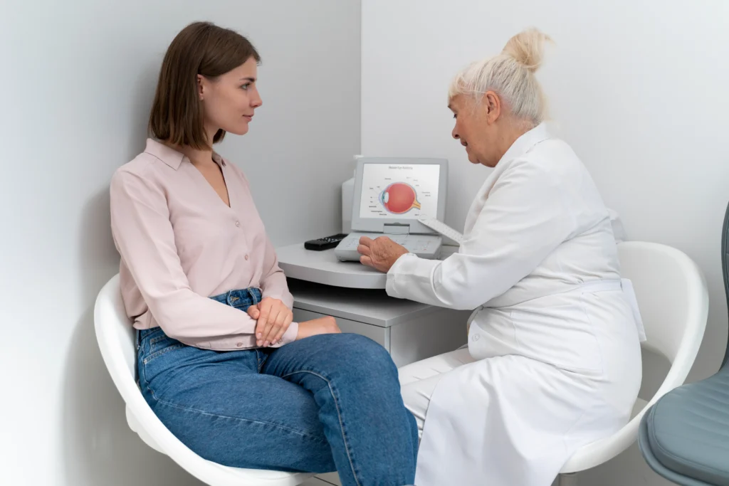
The basic principle is similar to sonar, the technology used by submarines and marine vessels. Sound waves are sent into the body, they bounce off internal structures, and the returning echoes are converted into a visual image on a screen. A trained provider reads those images to evaluate organs, identify abnormalities, and decide on next steps in your care.
Here is a plain-language breakdown of what actually happens during an ultrasound examination:
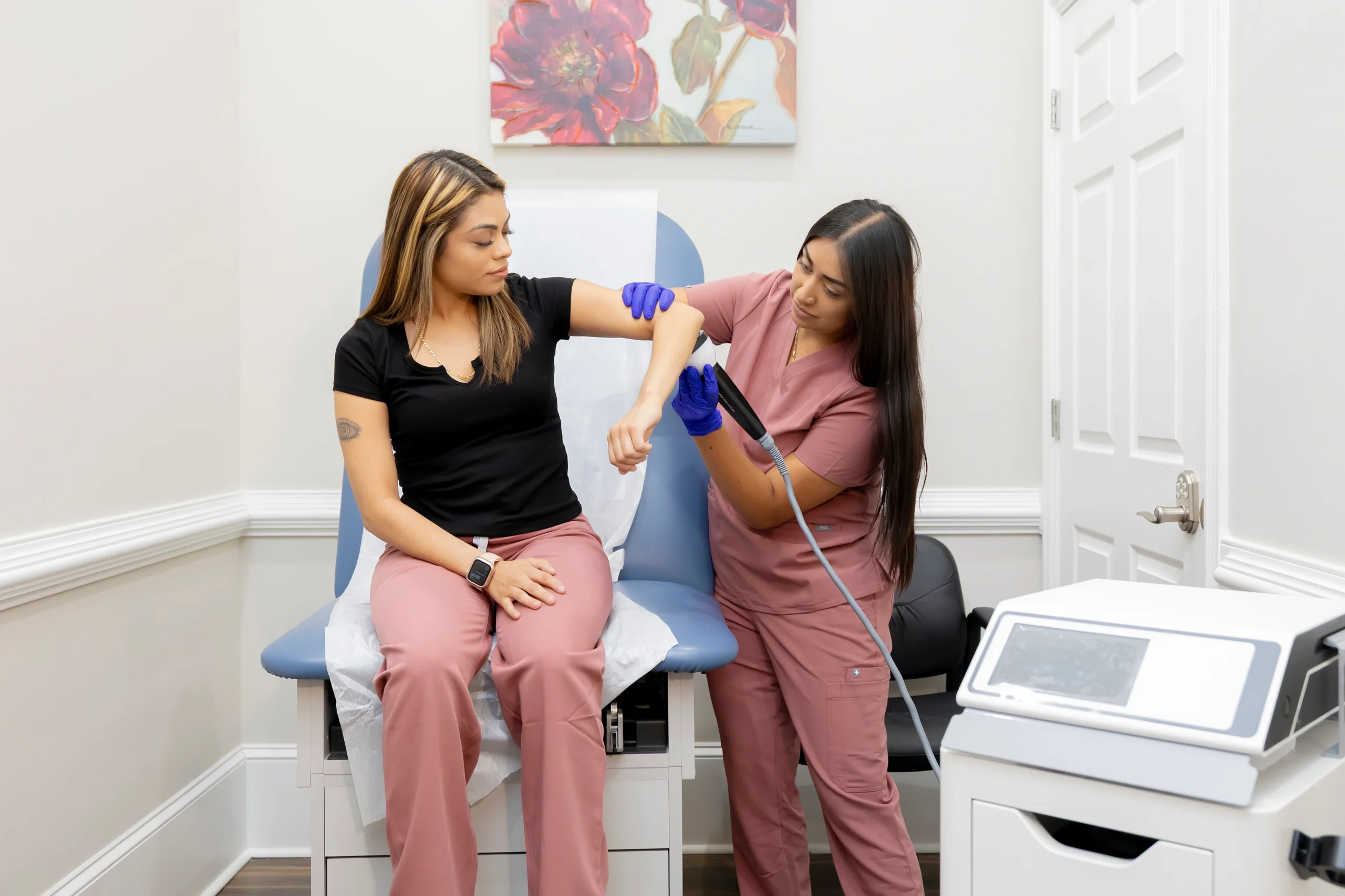
Before the exam starts, a water-based gel is applied to the skin over the area being scanned. The gel eliminates air pockets between the transducer and the skin that would scatter the sound waves and reduce image quality. It wipes off easily when the exam is finished. The examination itself is completely painless.
This is the most common type. It produces two-dimensional cross-sectional images of internal structures and is used to evaluate abdominal organs, the thyroid, soft tissue masses, and many other areas of the body.
Doppler ultrasound measures the speed and direction of blood flow within vessels. It is essential for vascular studies and for detecting conditions like blood clots or arterial narrowing.
An advanced form of Doppler that color-codes blood flow based on its direction, typically showing flow moving toward the transducer in red and flow moving away in blue. It is widely used in cardiovascular and vascular assessments and makes it much easier to spot abnormal flow patterns at a glance.
M-mode displays the movement of structures over time along a single axis. It is commonly used in echocardiography to assess how heart valves are opening and closing and to measure cardiac dimensions.
Ultrasound is one of the most versatile diagnostic tools in clinical medicine. At Zam Zam Family Clinic, our providers use in-clinic ultrasound to evaluate conditions across multiple organ systems.
Patients sometimes wonder why their provider ordered an ultrasound rather than an X-ray, CT scan, or MRI. Here is how they actually differ in practice.
Ultrasound vs X-ray: X-rays are excellent for imaging bone and detecting fractures, but they provide limited information about soft tissue and they use radiation. Ultrasound is better for evaluating organs, blood vessels, and soft tissue, and it carries zero radiation exposure.
Ultrasound vs CT scan: CT scans offer highly detailed cross-sectional images and are invaluable for complex anatomy, trauma assessment, and cancer staging. They use ionizing radiation and take longer to complete. Ultrasound is faster, portable, radiation-free, and ideal for initial screening or when real-time imaging is needed.
Ultrasound vs MRI: MRI provides the best detail for soft tissue structures, particularly the brain, spine, and joints. It is also significantly more expensive, requires the patient to remain completely still inside a large machine for 30 to 60 minutes, and is not appropriate for patients with certain metal implants. Ultrasound is far more accessible, faster, and well suited for most first-line evaluations.
Ultrasound is often the first imaging step precisely because it is safe, affordable, widely available, and gives providers immediate, actionable information. It frequently guides the decision about whether more advanced imaging is even necessary, and if so, what type would be most useful.
Yes, without reservation. Diagnostic ultrasound has been in clinical use for more than six decades and has an outstanding safety record. At diagnostic intensities, sound waves have no known harmful biological effects. There is no radiation involved, no contrast dye required in the vast majority of exams, and no recovery period after the scan.
Unlike X-rays, CT scans, or nuclear imaging, there is no cumulative exposure concern with ultrasound. Patients who need ongoing monitoring over months or years can receive ultrasound exams as frequently as their clinical situation requires, without any radiation-related risk to consider.
Having ultrasound imaging available directly at your primary care clinic changes the experience in a very practical way. You do not need to schedule a separate appointment at an imaging center, find parking at an unfamiliar location, or wait for records to travel between offices. The scan happens in the same place where your provider already knows your full medical history.
At Zam Zam Family Clinic, we have intentionally built a practice where patients can access the diagnostics they need without being sent elsewhere for basic imaging. In practical terms, that means:
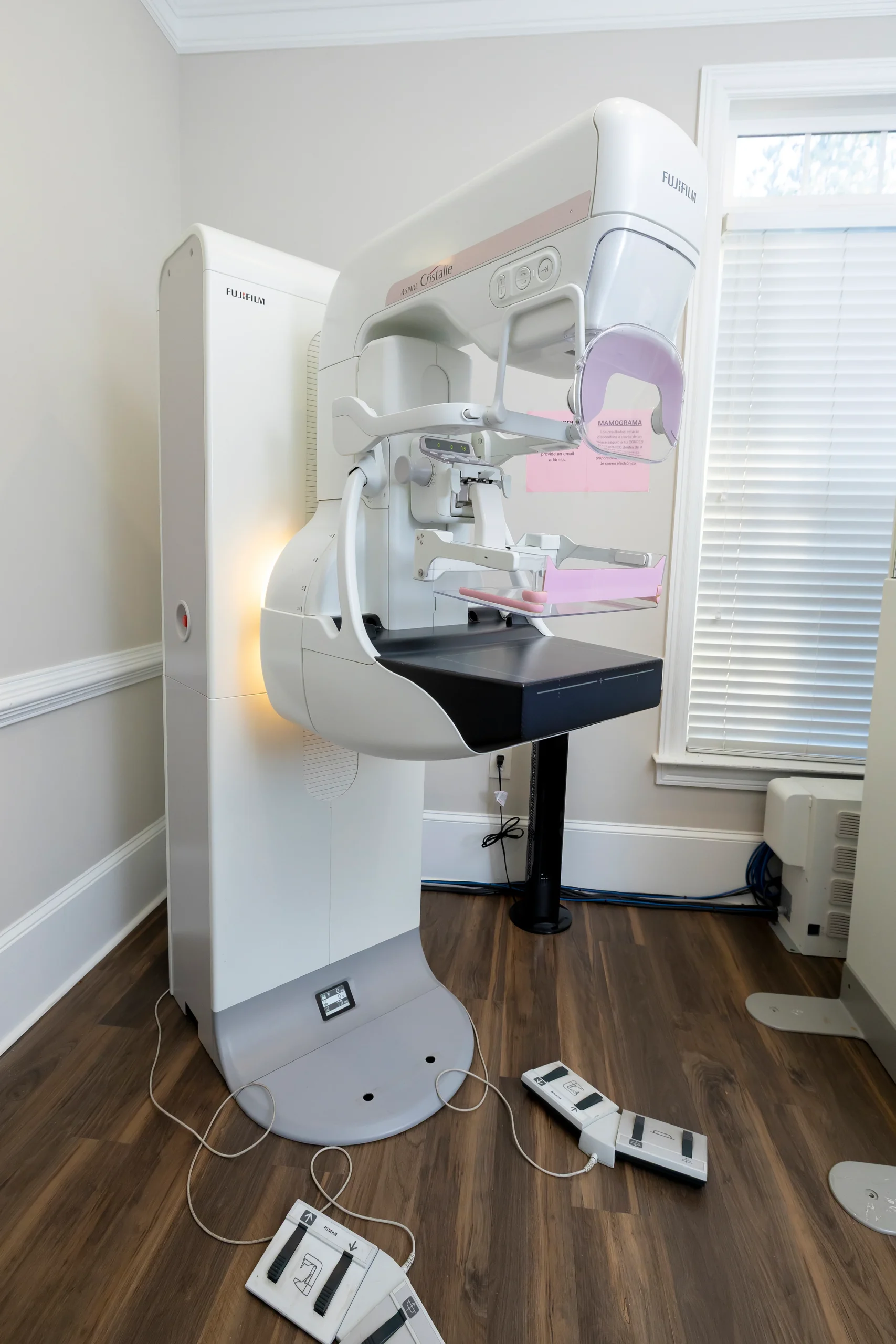
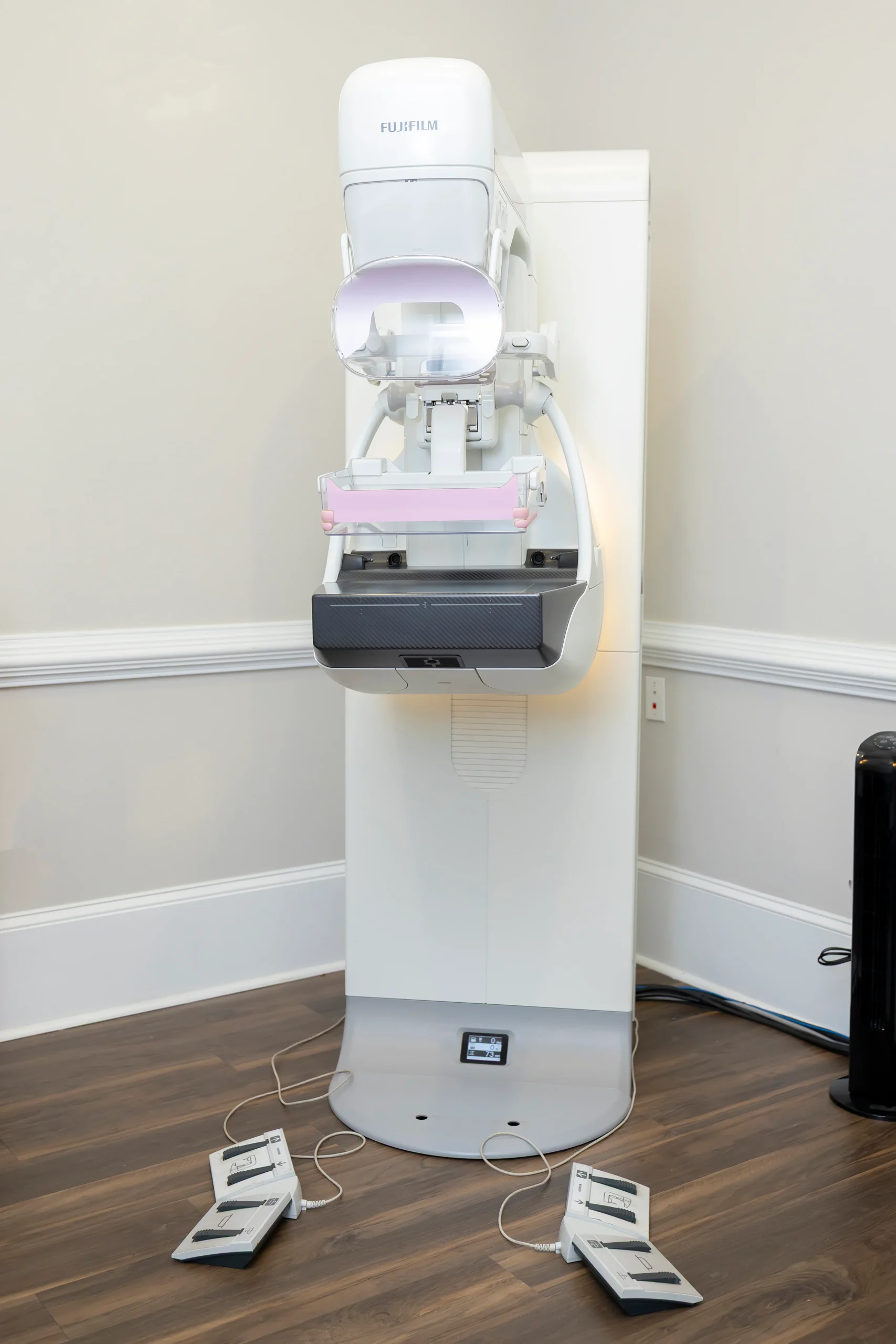
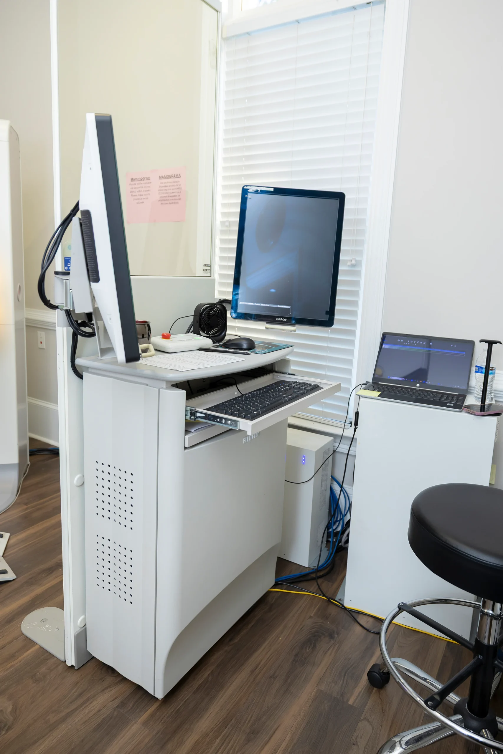
Our in-clinic imaging capabilities include ultrasound, mammography, echocardiogram, and vascular and cardiovascular testing, all at 965 Oakland Road in Lawrenceville, GA 30044.
Mon - Fri: 8:00 am – 5:00 pm
Sat & Sun: Closed
Terms & Conditions | Privacy Policy | Sitemap